


Annabel Ensor BVSc.
Ringworm
Ringworm is a common contagious skin disease of horses. It is caused by a fungal infection. Trichophyton equinum, Trichophyton mentagrophytes, Microsporum gypseum, and Microsporum canis are the most frequently isolated organisms. It is important to note that humans can become infected by handling infected horses and contaminated tack.
Infection is transmitted among horses by direct contact with an infected animal (including humans) or by indirect contact via contaminated tack, covers, or fungal spores within the environment.
Illness, poor nutrition, overcrowding, age and stressful environments predispose horses to infection. Young and elderly horses are more susceptible to infection. Continuous wetting of the horse’s skin e.g. sweating, washing-down can decrease the skin’s protective barrier therefore enabling infection to occur.
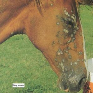
The classic ringworm lesion is a circular patch of hair loss with stubbly hairs on the margin. The skin can appear to be crusted and/or scaly. Ringworm lesions usually begin as small raised lumps that progress to the typical ringworm lesion. Some horses can become itchy and may also show evidence of pain.
Ringworm is usually a self-limiting disease and most horses recover within 1-6 months. Treatment is usually advisable as it decreases the transmission of disease between horses and other animals including humans. Antifungal shampoos (e.g. ‘Three Horses’ fungal shampoo) and/or washes e.g. 3% Captan, povidone iodine and lime sulphur, are useful for decreasing the transmission of disease but do not alter the course of infection. Systemic treatment with antifungal drugs is effective but this is very expensive and it’s use is usually limited to humans and small animals e.g. dogs and cats.

Warts
Warts (equine viral papillomatosis) are caused by a type of virus known as papovavirus. Horses less than 3 years of age are most commonly affected. Transmission appears to be by direct contact between infected horses. Lesions are usually multiple and are most commonly found around the lips and nose but can also be present on the ears and legs.
Warts are best left untreated because spontaneous remission almost always occurs. They can be very unsightly therefore a variety of treatments have been devised to improve the horse’s cosmetic appearance.

Rain Scald
Rain scald (dermatophilosis) is an infection caused by the bacterium Dermatophilus congolensis. This organism also causes mud-fever. The source of the organism is thought to be carrier animals, scabs from infected animals and/or soil. Infection requires prolonged exposure to moisture and damaged skin.
- Remove all scabs
- Apply a topical antibacterial solution e.g. chlorhexidine, povidone iodine
- Keep the affected area dry
Sarcoids
Sarcoids are the most common skin tumour in horses. They are classified as benign but can invade local tissues. Benign tumours do not spread throughout the body. Sarcoids are caused by a yet to be identified agent, some researchers believe that a virus is involved. Sarcoids often develop in areas that were previously injured. They are common in young horses (less than 4 years of age). The most commonly affected areas include the head, ears and legs. Your veterinarian can confirm the diagnosis of a sarcoid by carrying out a biopsy.There are four types of sarcoid:
Flat
These can resemble ringworm, rain scald, and bacterial skin infections. They appear as circular areas of hair loss with scaling and crusting and may extend deep into the tissues.

Fibroblastic (proliferative)
These appear as nodules or larger ulcerated masses. They are similar to proud flesh.

Verrucous (wart-like)
These are wart-like in appearance and rarely grow greater than 6cm in diameter.

A mixture of verrucous and fibroblastic
Removing and preventing the recurrence of sarcoids can be quite challenging for the veterinarian. Several treatment options are available and these often depend on the location and type of the sarcoid(s) that are present.
Some of the treatments that are available include:
- “Watchful neglect” is often a feasible approach if there is a single non-changing lesion and no evidence of infection.
- Cryosurgery involves freezing the mass. The sarcoid is destroyed by ice crystals that form inside the cells, rupturing the cell walls and killing the tissue. It can take greater than 8 weeks for complete healing to occur.
- Excision-removing the mass surgically. Generally, this is relatively quick, inexpensive and does not require repeated veterinary visits. Large margins surrounding the sarcoid need to be taken, otherwise the sarcoid can reoccur. Surgical excision is difficult on the lower limbs and face, because the skin is tighter, healing is slower and there is an increased chance of scarring. Also, large margins cannot be taken therefore there is an increased chance of the sarcoid returning.
- Intralesional injection of bacilli Calmette-Guerin vaccine (BCG), this stimulates the horse’s immune system to recognise the sarcoid as foreign and thus destroy it. Multiple treatments are often required. This treatment is especially useful in areas in which it is difficult to surgically remove the sarcoid e.g. the eyelid
- ChemotherapyAn anti-cancer agent is injected into the tumour site. The primary aim is to stop cell division (reproduction) and thus cause cell death. Multiple injections are required. Cisplatin and 5-fluorouracil are the most commonly used chemotherapeutic agents. This treatment can be expensive due to the cost of the drugs.